Swollen and Erythematous Finger Following a Paper Cut
AUTHORS:
Joanna Theophilopoulos, BS1; Keval Patel, BS1; Kathryn E. Wheeler, MD2; Kathleen A. Ryan, MD2; Carolyn G. Carter, MD, MS2
1University of Florida College of Medicine
2Department of Pediatrics, University of Florida College of Medicine
Case Report | PUBLISHED FALL 2022 | Volume 42, Issue 4
DOWNLOAD PDF
Case Report
A 5-year-old girl with an unremarkable medical history presented to her pediatrician with swelling and blistering of her left index finger. Ten days prior, she sustained a paper cut on the palmar surface of the middle phalanx of this finger. Over the next few days, she swam at the beach and in a river. Subsequently, the wound became erythematous and swollen. Approximately five days after the paper cut, blisters began to erupt. The patient was taken to an urgent care clinic where she was prescribed cephalexin for a presumed bacterial infection.
Over the next 48 hours, the phalanx became increasingly erythematous and more blisters erupted, which prompted a visit to her pediatrician. On exam, there was erythema and edema of the middle phalanx of the left index finger with a healing laceration on the palmar surface surrounded by multiple clear, fluid-filled vesicles (Figure 1). She had mobility of the finger, but it was painful. The parents denied any drainage from the vesicles or systemic symptoms. Two of the vesicles were punctured and fluid was sent for bacterial culture and HSV PCR. The patient’s antibiotic regimen was changed from cephalexin to clindamycin and topical mupirocin.
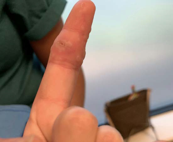
Figure 1
Three days later, the patient presented with worsening symptoms, despite using the antibiotics as prescribed. On exam, there was increased blistering of the left index finger, as well as an additional blister on the center of her left palm (Figure 2). The erythema and edema persisted, and the patient had mildly limited active range of motion. She denied any pain, tingling, or itching. At this point, the parents informed the pediatrician the patient’s sister recently had resolution of cold sores prior to the patient developing blisters on her finger. During this visit, the bacterial culture came back negative, but the HSV PCR was still pending. The patient was prescribed oral acyclovir 400 mg three times daily for five days awaiting the PCR results, and clindamycin was discontinued.
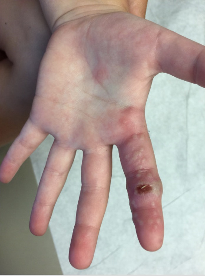
Figure 2
Two days later, the patient returned to the urgent care clinic due to development of additional lesions that appeared pustule-like. The urgent care physician suspected MRSA and resumed oral clindamycin, in addition to continuing acyclovir and mupirocin.
The patient was referred to the Pediatric Infectious Disease Clinic for further evaluation the following day. The blisters turned cloudy, with reddened edges, and there were two new lesions on the palm. The lesions had not spread anywhere else on the body, and she had no fever, malaise, or headache. She still had functional use of her hand, but she reported tactile pain. At this time, the HSV PCR from the initial visit to the pediatrician came back positive for HSV-1 and negative for HSV-2. The patient was diagnosed with herpetic whitlow.
Of note, the patient did not have a personal history of cold sores. However, her 12-year-old sister had such a history and recently recovered from an oral lesion. The patient’s father also had a distant history of cold sores. Since the patient had no history of HSV but was in very close contact with her sister who recently had a lesion, this was likely the source of the exposure. The patient’s herpetic whitlow was likely a primary HSV infection, accounting for the severity of the lesions.
At the infectious disease clinic, the lesions appeared to be healing. The mother reported that no new lesions had appeared for the preceding few days. Due to ongoing concern for a bacterial superinfection, a bacterial culture of the vesicular fluid was repeated. The fluid was clear in appearance. Five days later, the repeat bacterial culture came back negative. The patient’s mother reported significant improvement at this point with minimal swelling. No blisters remained and the skin was desquamating where the blisters had been. At this point clindamycin was discontinued, and the patient completed the 10-day course of acyclovir.
Discussion
Herpetic whitlow is an infection of the phalanx or hand caused by Herpes Simplex Virus Type 1 or 2 (HSV1 or HSV2). HSV is spread via direct contact of mucous membranes or broken epidermis.1, 2 Herpetic whitlow can result if broken epidermis on the hand is exposed to either oral or genital herpetic lesions. In adults, herpetic whitlow often occurs via exogenous inoculation. Healthcare workers, such as dentists, can be affected if they are exposed to secretions from patients with oral herpetic lesions.3 In children, herpetic whitlow typically occurs via autoinoculation when a child has coexisting gingivostomatitis or herpes labialis. However, there have been several cases of children contracting herpetic whitlow without a personal history of HSV.4–7 Exogenous inoculation from close contacts who are infected can also occur in children.6, 7 This is likely what occurred in the case we have presented, as the parents recalled the sister with the cold sore kissed the patient’s paper cut.
Herpetic whitlow lesions typically appear 2-20 days following inoculation.1, 2, 7 The lesions present as painful, non-purulent, fluid-filled vesicles on an erythematous base. After about a week, the fluid can become cloudy, which can mislead clinicians.6 In one case series, 65% of patients with herpetic whitlow were initially misdiagnosed with a bacterial infection.6 This case is unique in that the patient had a laceration associated with her herpetic whitlow. In addition, her exposure to salt and freshwater expanded the differential to water-borne pathogens. Furthermore, the patient’s lesions began to appear pustule-like several days after the blisters initially erupted. This was concerning for a superimposed bacterial infection but could have simply represented a natural progression of the lesions.
Understanding the natural progression of the lesions can help clinicians properly diagnose the infection and avoid unnecessary antibiotics. Typically, over the course of two weeks, the vesicles will crust and desquamate.7 Left untreated, herpetic whitlow typically resolves after 3-4 weeks.7 The skin will usually re-epithelialize without scars or deformities.7
Herpetic whitlow can have varying presentations in different patients. It can present as a single vesicle or in clusters. In some cases, there is associated regional lymphadenopathy or lymphangitis.8 There are rarely systemic symptoms, but in some cases, patients report a flu-like prodrome preceding the cutaneous symptoms.2 The most common prodromal symptoms are pain, tingling, or itching in the affected area.8
Diagnosis of herpetic whitlow can be made on clinical grounds, especially if the infection occurs in an individual with a history of HSV, or if the individual was in close contact with another person infected with HSV.6 Laboratory analysis can aid in making the diagnosis in more complex cases. Viral cultures and Tzanck smears have fallen out of favor due to limited sensitivity.7, 9 PCR testing is a much more sensitive option and can be done with the fluid obtained from a simple needle puncture of a vesicle.6, 9
Guidelines for treating herpetic whitlow are not well-established. In healthy patients, the infection is typically self-limiting, so treatment is not always necessary. Systemic acyclovir may shorten the course of the infection and prevent the eruption of additional vesicles.6 Treatment during prodromal symptoms may prevent eruption of blisters.10 The use of topical acyclovir is controversial. In one review of several cases, there was no reported benefit of topical therapy.6 However, more research is needed as some clinicians report anecdotal benefits.10
HSV remains latent in sensory ganglia after initial infection. Recurrence can be triggered by physiological and psychological stressors.1 Herpetic whitlow recurrence is observed in 20-50% of patients.1 Recurrent infections make up the majority of HSV hand infections and present more often in adults aged 20-40.11 Recurrence is typically preceded by prodromal symptoms including pain, tenderness, pruritus, burning, and aching.11 Prodromal symptoms vary in duration, ranging from 3 hours to 3 days. Primary infections are typically more severe than subsequent recurrent infections.12 There are no clear guidelines that address the management of recurrent herpetic whitlow. However, in a study of patients with recurrent herpetic whitlow caused by HSV-2, oral acyclovir (2 g/day in three doses for 10 days) started during the earliest phase of a recurrence resulted in a reduced duration of symptoms from 10.1 to 3.7 days.11
In healthy individuals, herpetic whitlow is typically a self-limiting infection. However, there are complications that should be considered. Superinfections with Staphylococcus aureus and other bacteria are the most common complication.6 Other complications include nail dystrophy and permanent nail loss.6 Rarely, herpetic whitlow can result in HSV encephalitis or meningitis.6, 13 Incision and drainage is contraindicated in herpetic whitlow, as this can increase the risk of viremia and superimposed bacterial infections.6, 8, 14 These complications illustrate the need for prompt and accurate diagnosis of herpetic whitlow.
Conclusion
Herpetic Whitlow is usually a self-limiting HSV infection of the hand or digits. Maintaining an index of suspicion and understanding the natural progression of the lesions are critical in diagnosing this infection. Our patient presented with a primary HSV-1 infection of her finger from familial inoculation of a superficial laceration. Her lesions spread and the vesicles became cloudy over a two-week period. Lack of improvement with antibiotic therapy and two negative bacterial cultures point away from a secondary bacterial infection as a cause for her worsening symptoms. She has recovered from her infection without apparent sequelae.
References
- Rubright JH, Shafritz AB. The herpetic whitlow. J Hand Surg Am. 2011 Feb;36(2):340-2. doi: 10.1016/j.jhsa.2010.10.014. Epub 2010 Dec 24. PMID: 21186084.
- Raykova VV. Learning from Mistakes – a Case of Pediatric Patient with Recurrent Whitlow. Folia Med (Plovdiv). 2019 Sep 30;61(3):478-480. doi: 10.3897/folmed.61.e39163. PMID: 32337938.
- Browning WD, McCarthy JP. A case series: herpes simplex virus as an occupational hazard. J Esthet Restor Dent. 2012 Feb;24(1):61-6. doi: 10.1111/j.1708-8240.2011.00469.x. Epub 2011 Aug 30. PMID: 22296698; PMCID: PMC3437498.
- Murphy AP, Martin P, Jukka CM, Memon A, Ng SM. Recurrent primary paediatric herpetic whitlow of the big toe. BMJ Case Rep. 2013 Feb 21;2013:bcr2013008598. doi: 10.1136/bcr-2013-008598. PMID: 23436893; PMCID: PMC3604391.
- Kopriva F. Recurrent herpetic whitlow in an immune competent girl without vesicular lesions. Eur J Pediatr. 2002 Feb;161(2):120-1. doi: 10.1007/s00431-001-0879-3. PMID: 11954749.
- Szinnai G, Schaad UB, Heininger U. Multiple herpetic whitlow lesions in a 4-year-old girl: case report and review of the literature. Eur J Pediatr. 2001 Sep;160(9):528-33. doi: 10.1007/s004310100800. PMID: 11585074.
- Walker LG, Simmons BP, Lovallo JL. Pediatric herpetic hand infections. J Hand Surg Am. 1990 Jan;15(1):176-80. doi: 10.1016/s0363-5023(09)91128-5. PMID: 2153722.
- Lieberman L, Castro D, Bhatt A, Guyer F. Case report: palmar herpetic whitlow and forearm lymphangitis in a 10-year-old female. BMC Pediatr. 2019 Nov 21;19(1):450. doi: 10.1186/s12887-019-1828-5. PMID: 31752766; PMCID: PMC6868856.
- Betz D, Fane K. Herpetic Whitlow. 2021 Aug 6. In: StatPearls [Internet]. Treasure Island (FL): StatPearls Publishing; 2021 Jan–. PMID: 29494001.
- Patel R, Kumar H, More B, Patricolo M. Paediatric recurrent herpetic whitlow. BMJ Case Rep. 2013 Jul 31;2013:bcr2013010207. doi: 10.1136/bcr-2013-010207. PMID: 23904423; PMCID: PMC3736177.
- Gill MJ, Bryant HE. Oral acyclovir therapy of recurrent herpes simplex virus type 2 infection of the hand. Antimicrob Agents Chemother. 1991 Feb;35(2):382-3. doi: 10.1128/AAC.35.2.382. PMID: 1850971; PMCID: PMC245012.
- Merchant VA, Molinari JA, Sabes WR. Herpetic whitlow: report of a case with multiple recurrences. Oral Surg Oral Med Oral Pathol. 1983 Jun;55(6):568-71. doi: 10.1016/0030-4220(83)90372-9. PMID: 6576289.
- Karpathios T, Moustaki M, Yiallouros P, Sarifi F, Tzanakaki G, Fretzayas A. HSV-2 meningitis disseminated from a herpetic whitlow. Paediatr Int Child Health. 2012 May;32(2):121-2. doi: 10.1179/2046905511Y.0000000004. PMID: 22595224.
- Louis DS, Silva J Jr. Herpetic whitlow: herpetic infections of the digits. J Hand Surg Am. 1979 Jan;4(1):90-4. doi: 10.1016/s0363-5023(79)80113-6. PMID: 759512.