Neonatal Lupus Erythematosus: A Case Report of an Infant with Annular Rash
AUTHORS:
Anna Walls, BS1; Anna Abbott, MD2; Zachary Gohsman, MD2
1University of Florida College of Medicine
2Department of Pediatrics, University of Florida College of Medicine
CASE REPORT | PUBLISHED Winter 2026 | Volume 46, Issue 1
DOWNLOAD PDF
Abstract
Neonatal lupus erythematosus is a rare autoimmune disorder caused by the transplacental passage of maternal autoantibodies, primarily anti-SSA/Ro and anti-SSB/La. It commonly presents with dermatologic manifestations and, less frequently, cardiac, hepatic, and hematologic involvement.1 We report the case of a 6-week-old male who presented with a widespread annular rash. Despite an unremarkable perinatal history, physical examination of the patient’s mother revealed that she had a facial rash raising strong suspicion for undiagnosed systemic lupus erythematosus. Laboratory testing of the patient confirmed elevated anti-SSA and anti-SSB antibodies, consistent with neonatal lupus erythematosus. The patient exhibited hematologic abnormalities, including neutropenia and anemia, but cardiac evaluation, including ECG, Holter monitoring, and echocardiography, showed no conduction defects. The infant’s rash gradually resolved, consistent with the self-limiting nature of dermatologic neonatal lupus erythematosus. This case underscores the diagnostic challenges of neonatal lupus erythematosus, particularly in infants born to asymptomatic mothers. It highlights the importance of considering maternal autoimmune disease in cases of unexplained neonatal rashes. Given that many affected mothers lack a prior diagnosis of autoimmunity, maternal antibody screening during pregnancy may aid in early identification of at-risk neonates. While dermatologic findings typically resolve as maternal antibodies degrade, cardiac involvement can have significant long-term consequences, necessitating close monitoring and multidisciplinary care. This case emphasizes the need for heightened clinical suspicion, prompt recognition, and appropriate follow-up to optimize neonatal outcomes and identify undiagnosed maternal autoimmune conditions.
Background
Neonatal lupus erythematosus (NLE) is an acquired autoimmune condition resulting from the transplacental passage of maternal autoantibodies, specifically anti-Sjögren’s-syndrome-related antigen A (anti-SSA/Ro), anti-Sjögren’s-syndrome-related antigen B (anti-SSB/La), or anti-U1 ribonucleoprotein (anti-U1-RNP). It is a rare disorder, with an estimated incidence of 1 in 20,000 live births in the United States.1
Primary Objective
This report aims to illustrate the importance of considering NLE in infants with unexplained annular rash, even in the absence of known maternal autoimmune disease, to facilitate early diagnosis and appropriate management.
Subject Presentation
We present the case of a 6-week-old male born at full term (40 weeks and 5 days) to a 37-year-old G2P2 mother via normal vaginal delivery, with APGAR scores of 9 at both 1 and 5 minutes. The infant’s medical history was unremarkable until he presented to his pediatrician with a one-week history of rash. The rash initially appeared on the face and later spread to the head, arms, trunk, legs, and diaper area. The lesions remained unchanged since their onset. The patient had no history of fever or upper respiratory symptoms. The mother was breastfeeding and supplementing with formula. She reported switching the formula about a week before the rash developed, but no other changes in the patient’s oral intake were noted. No other household members had a similar rash.
At presentation, the infant was afebrile, with a height and weight in the 84th and 57th percentiles, respectively. Examination revealed a widespread erythematous, blanching rash involving the face, head, trunk, legs, and groin. The rash consisted of annular lesions with maculopapular features, flat lesions on the trunk and extremities (Figures 1 and 2), and slightly raised lesions on the head (Figure 3).
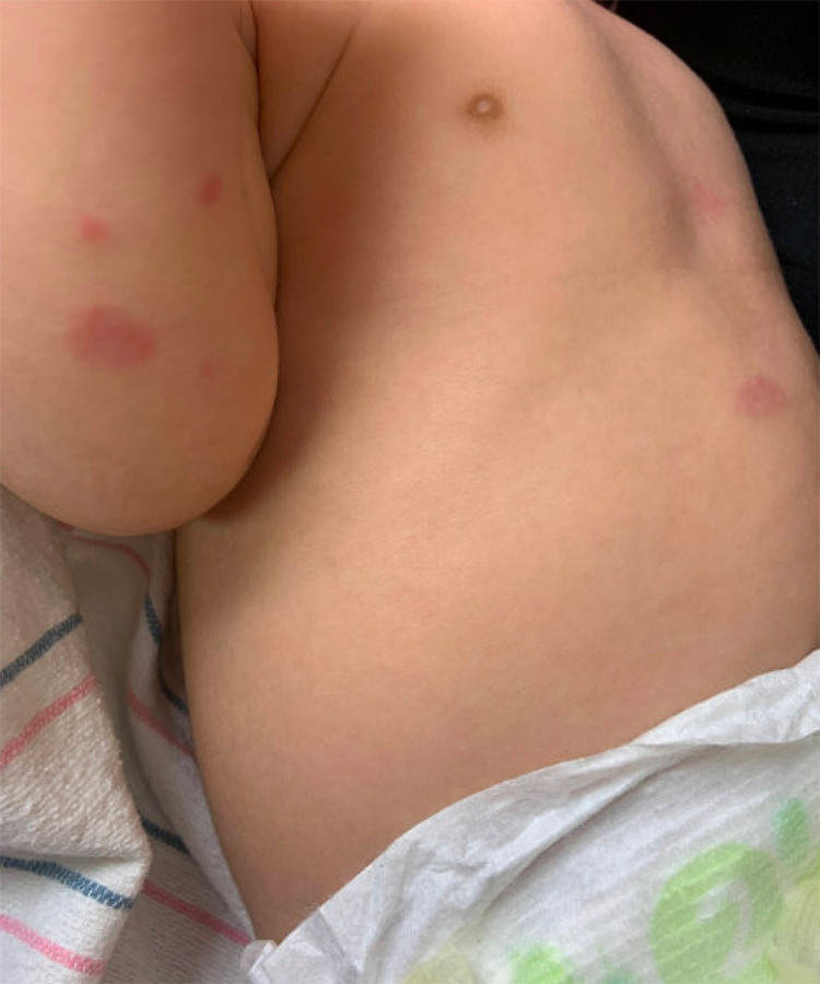
Figure 1 (see text)
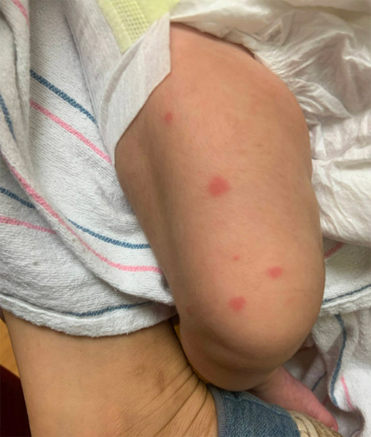
Figure 2 (see text)
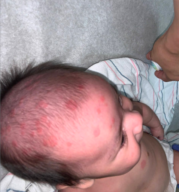
Figure 3 (see text)
The appearance of the rash prompted the clinician to investigate the mother’s medical history further, revealing that she had an undiagnosed facial rash since 2014 (Figure 4) and a history of carpal tunnel syndrome. Neonatal lupus emerged as the leading differential diagnosis, prompting anti-SSA/Ro and anti-SSB/La antibody testing. Other differentials were considered, including papular eczema and scabies/mite infestation. However, scabies/mites were deemed less likely due to the absence of symptoms in other family members and the lack of resolution in the lesions.
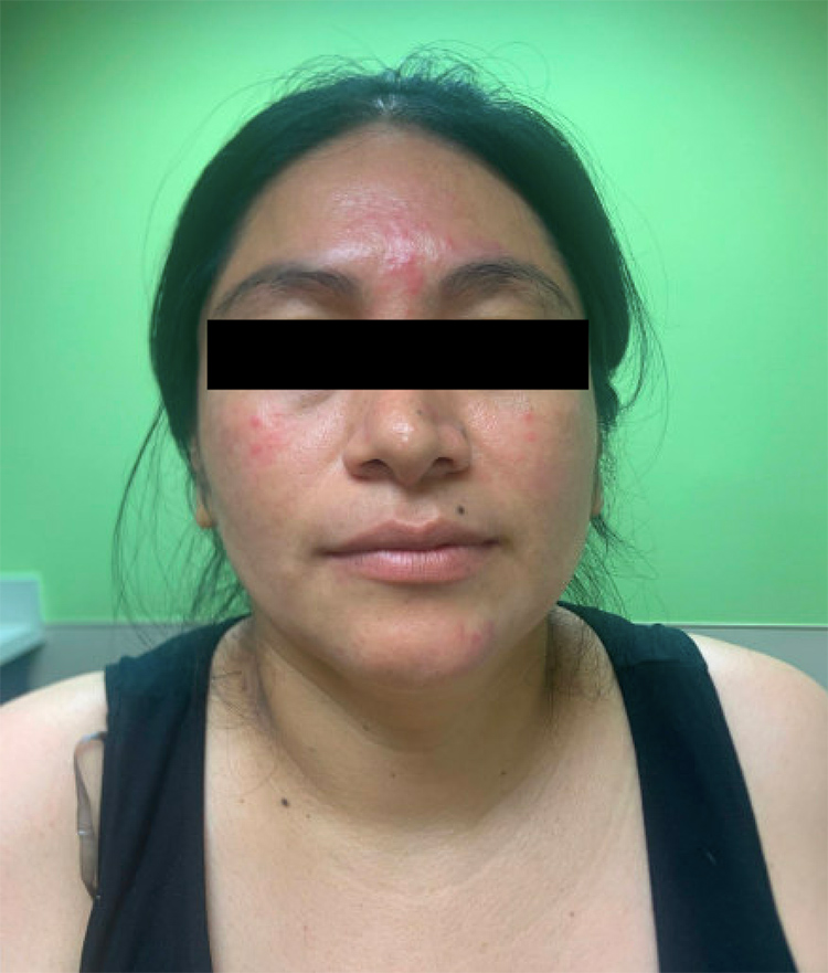
Figure 4 (see text)
Laboratory testing confirmed the diagnosis of NLE with elevated anti-SSA antibodies (7.2 units/mL) and anti-SSB antibodies (>8 units/mL). The patient’s mother was informed of the diagnosis and returned to the clinic a few days later. At this visit, the patient’s rash was noted to be non-bothersome and showed signs of resolution, with no new lesions present. After consultation with pediatric rheumatology, the care plan included obtaining an EKG, repeating antibody testing when the patient reached 6 and 12 months of age, and referring the patient to cardiology and dermatology.
At the dermatology visit, additional confirmatory tests were ordered, including a complete blood count (CBC), a comprehensive metabolic panel (CMP), and several autoimmune markers. CBC showed a hemoglobin of 9.3 g/dL and neutropenia (absolute neutrophil count of 1.08 cells/µL, neutrophils 14%), but no lymphopenia and a normal white cell count and platelets. CMP showed an AST of 38U/L but otherwise normal liver and renal function. The patient’s antibody testing was positive for ANA (titer of 1:1280 and speckled pattern) and chromatin (67 AI) and negative for dsDNA (1 IU/mL), Sm/RNP (7 units/mL), Scl-70 (2 AU/mL), and Smith antibodies (3 AU/mL).
Cardiology evaluation included an EKG showing normal sinus rhythm without conduction abnormalities, a Holter monitor displaying sinus rhythm with occasional sinus tachycardia, and an echocardiogram showing normal anatomy and function. These tests confirmed that the patient did not exhibit any cardiac manifestations of NLE.
At the two-month well-child visit, the mother reported significant improvement in the infant’s rash, and the child continued to breastfeed well. The mother was subsequently tested and diagnosed with systemic lupus erythematosus (SLE).
Discussion
NLE is characterized by maternal autoantibodies crossing the placenta and affecting the fetus. Clinical manifestations include dermatologic, cardiac, hepatic, neurologic, and hematologic features. Cardiac manifestations, which are the most serious, include congenital heart block, myocarditis, and valvular dysplasia. One postulated mechanism of cardiac pathogenesis involves anti-Ro-mediated calcium dysregulation leading to apoptosis. Anti-La antibodies then form immune complexes with apoptotic cells, activating inflammatory cascades that disrupt cardiac conduction.2 Unlike dermatologic features, which typically resolve as maternal antibodies degrade, cardiac manifestations are irreversible and may lead to significant morbidity and mortality. Fortunately, infants with NLE presenting with only non-cardiac manifestations and no evidence of heart block at birth by examination and ECG are unlikely to develop cardiac disease.
This case underscores the importance of considering NLE in the differential diagnosis of infants presenting with rash, especially annular rash, even in the absence of a significant maternal autoimmune history. Studies have shown that many mothers of infants with NLE are asymptomatic at the time of delivery. One study reported that only one in 20 cardiac NLE cases occurred in a child born to a mother previously diagnosed with SLE.3 At the same time, another found that 64% of mothers of infants with NLE were asymptomatic.4 These findings highlight the diagnostic challenges and emphasize the potential value of screening for antinuclear antibodies during pregnancy to identify at-risk pregnancies.5
In conclusion, clinicians should maintain a high index of suspicion for NLE in infants presenting with unexplained rashes. Early recognition and multidisciplinary management are crucial to preventing complications and ensuring favorable outcomes.
Author Disclosure Statement: The authors report no conflicts of interest.
References
- University of Mansoura, Nasef N. Neonatal Lupus Erythematosus. Neonatol Clin Pediatr. 2014;1(1):1-10.
- Izmirly P, Saxena A, Buyon JP. Progress in the pathogenesis and treatment of cardiac manifestations of neonatal lupus. Curr Opin Rheumatol. 2017;29(5):467-472.
- Skog A, Lagnefeldt L, Conner P, et al. Outcome in 212 anti-Ro/SSA-positive pregnancies and population-based incidence of congenital heart block. Acta Obstet Gynecol Scand. 2016;95(1):98-105.
- Wisuthsarewong W, Soongswang J, Chantorn R. Neonatal lupus erythematosus: clinical character, investigation, and outcome. Pediatr Dermatol. 2011;28(2):115-121.
- Derdulska JM, Rudnicka L, Szykut-Badaczewska A, et al. Neonatal lupus erythematosus – practical guidelines. J Perinat Med. 2021;49(5):529-538.