
Chronic Nonbacterial Osteomyelitis in Pediatric Inflammatory Bowel Disease: A Case Series
AUTHORS:
Natalie Jennings, DO; Monica Ramirez, MD; Elizabeth Torres Claudio, MD; Lina Felipez, MD
Nicklaus Children’s Hospital, Miami, FL
Case report | PUBLISHED Fall 2026 | Volume 46, Issue 4
DOWNLOAD PDF
Abstract
Objectives/Background
Chronic non-bacterial osteomyelitis is a rare, sterile bone auto-inflammatory disorder that mainly affects children and adolescents. It can present as an extraintestinal manifestation of inflammatory bowel disease. We present a case series of 12 pediatric inflammatory bowel disease patients diagnosed with chronic non-bacterial osteomyelitis in our tertiary care center in South Florida, analyzing their demographics, symptoms, and imaging findings to explore the association between chronic non-bacterial osteomyelitis and inflammatory bowel diseas
Methods
We retrospectively reviewed the electronic medical records of pediatric inflammatory bowel disease patients diagnosed with chronic non-bacterial osteomyelitis at our institution between 2011 to 2024, assessing demographic data, clinical features, and imaging findings
Results
Of the 12 patients, 6 had Crohn’s disease, and 6 had ulcerative colitis. The median age of inflammatory bowel disease onset was 11.7 years, with chronic non-bacterial osteomyelitis onset occurring at a median age of 13.3 years and a median diagnostic interval of 1.75 years. CNO was diagnosed after inflammatory bowel disease in 8/12 (67%), before inflammatory bowel disease in 1/12 (8%), and concurrently in 3/12 (25%) of patients. Chronic non-bacterial osteomyelitis symptoms included multifocal pain (5/12, 42%), swelling (2/12, 17%), and limping (2/12, 17%), with incidental findings in 5/12 patients (42%). MRI showed multifocal lesions in 9/12 (75%), with axial skeleton involvement most common (7/12, 58%)
Conclusion
Our 13-year case series supports the association between chronic non-bacterial osteomyelitis and inflammatory bowel disease, with most patients developing chronic non-bacterial osteomyelitis after inflammatory bowel disease diagnosis. While multifocal pain was the most common symptom, some cases were asymptomatic, highlighting the role of imaging in early detection.
Introduction
Chronic non-bacterial osteomyelitis (CNO) is a noninfectious auto-inflammatory bone disease that primarily affects children and adolescents. The presentation varies, ranging from mild, isolated, self-limiting lesions to chronic and/or recurrent multifocal disease.1 The more severe presentation of CNO is referred to as chronic recurrent multifocal osteomyelitis (CRMO).2 CNO or CRMO most commonly affects females, with an average age of disease onset of 10 years.3 It has not been reported to be associated with a certain ethnicity. Because CNO/CRMO has nonspecific clinical, laboratory, and radiographic findings, delayed diagnosis is common. A retrospective survey of 284 patients performed in the U.S.A. reported a median duration from first symptoms to diagnosis of two years. However, with increased awareness, median time to diagnosis has decreased over time.4 The annual incidence rate has increased from approximately 0.4 per 100,000 children in 2010 to approximately 2.3 per 100,000 in 2019.5
The clinical presentation of CNO/CRMO is characterized by chronic waxing and waning bone pain. This may include nocturnal bone pain, which can mislead physicians to believe it to be due to other causes such as growing pains or bone tumors.1 Although lesions may occur in any bone, they are most commonly found in the metaphysis of the lower extremity long bones, as well as the clavicle, vertebra, pelvis, mandible, and long bones of the upper extremities. Patients may also have swelling over bones with little overlying soft tissue and experience fever, night sweats, and/or weight loss, further complicating the clinical picture. Other manifestations may include signs of inflammation in other organs, such as inflammatory bowel disease (IBD), psoriasis, palmoplantar pustulosis, and severe acne.2 CNO/CRMO is a diagnosis of exclusion, so patients oftentimes have a broad diagnostic workup before coming to the final diagnosis. Because clinicians may initially suspect bacterial osteomyelitis, patients often undergo lab workups that include a complete blood count (CBC), C-reactive protein (CRP), and erythrocyte sedimentation rate (ESR), which are typically normal or slightly elevated. Malignancy is also common in the differential; therefore, blood uric acid and lactate dehydrogenase levels are typically collected, and biopsies are performed, which usually show nonspecific findings revealing fibrosis, immune cells, bone lysis, and/or normal bone.1 Vitamin C levels may be obtained to test for scurvy, which can present with vague bone pain and limping or refusal to walk.
Goldfarb et al. discussed a case of a 5-year-old male who initially presented with symptoms concerning for scurvy and was later found to have CNO, and endoscopy/colonoscopy confirmed underlying Crohn’s disease.6 Although uncommon, CNO can be an extraintestinal manifestation of patients with IBD and may even explain arthralgia and bone pain in some IBD patients.7 Lorenze and colleagues described a case where a 10-year-old patient initially presented with signs of Crohn’s Disease that was diagnosed via colonoscopy, then presented two years later with vague bone pain and fevers that only improved with the initiation of steroids.8
Currently, no confirmatory diagnostic test exists for CNO/CRMO, and it remains primarily a diagnosis of exclusion. Initial diagnostic imaging often includes plain radiographs, which may be normal or reveal sclerotic, lytic, or mixed lesions depending on disease stage.9 However, isolated bone edema in the absence of structural damage will be missed on plain films.10 Whole-body magnetic resonance imaging (WB-MRI) remains the gold standard.11 It is also recommended to obtain WB-MRI rather than localized MRI to identify clinically silent lesions. The use of conventional computer tomography or ultrasound for diagnosing CNO is diminishing with improved knowledge of the condition; however, these methods may be useful in the initial stages of diagnosis to rule out other causes, such as malignancy, and to look for soft-tissue pathology.10
No gold-standard regimen currently exists for patients with this disease. Oftentimes, non-steroidal anti-inflammatory drugs (NSAIDs) are first-line therapy, having both anti-inflammatory and analgesic effects. NSAIDs control prostaglandin E production by inhibiting cyclooxygenases, thereby affecting nociception. Prostaglandin E is also essential for osteoclast activity; therefore, NSAIDs may inhibit osteoclasts and reduce pathological bone remodeling.10 Similarly, for their inflammation-reducing effects, corticosteroids have been used as either singular treatments or as a bridging agent when disease-modifying anti-rheumatic drugs (DMARDs) are used. In addition, anti-TNF agents and bisphosphonates have also been documented. Many of these treatment regimens are the same as medications used in pediatric IBD. The optimal treatment duration and when to discontinue therapy remain unclear, although complete resolution of lesions on MRI may indicate disease control.9 One of the most important aspects of treatment is monitoring for disease activity and relapse, which historically has been done via pain scores, routine inflammatory parameters, and imaging results via WB-MRI.2 Although prompt treatment with the correct agent is usually associated with favorable outcomes, there are risks for potential complications of CNO/CRMO. These may include growth/stature delay, limb-length discrepancy, and fractures. We conducted a retrospective chart review at our institution to identify the presentation and characteristics of CNO in conjunction with IBD to promote recognition of this disorder.
Methods
We conducted a single institutional retrospective chart review from 2011 to 2024 of pediatric patients aged 3-17 years at first diagnosis who presented to Nicklaus Children’s Hospital, a 309-bed tertiary care children’s hospital, and were diagnosed with both IBD and CNO/CRMO within the last 13 years. All patients were consented for the pediatric IBD research database with IRB approval. We searched the institutional electronic medical records software (CERNER) utilizing the International Classification of Diseases, Tenth Revision (ICD-10) diagnostic codes: Crohn’s disease (ICD-10 K50.9) or UC (ICD-10 K51.6) in combination with the diagnosis of chronic non-infectious osteomyelitis (ICD-10 M86.38).
The Jansson and Bristol criteria are both proposed diagnostic tools for CNO, although they have not been validated to date. The Jansson criteria use a scoring system with major and minor criteria such as multifocal bone lesions, elevated inflammatory markers, and response to NSAIDs to support diagnosis. In contrast, the Bristol criteria offer a more clinical approach, focusing on characteristic imaging findings, symptoms lasting over six weeks, lack of response to antibiotics, and exclusion of infection or malignancy.12,13 Because neither tool has been validated, our inclusion criteria focused on pediatric patients diagnosed with both IBD and radiographically confirmed CNO via MRI findings associated with active disease, such as bone edema, periosteal reaction, and soft tissue inflammation. There were no exclusion criteria. Two investigators independently reviewed the records and identified 12 unique patients who met these criteria. We analyzed their demographic factors, clinical features, imaging findings, and CNO treatment received.
Results
The study involved 12 participants, divided equally into two groups with Crohn’s disease (CD) and ulcerative colitis (UC), as shown in Table 1. Regarding race, 50% (6 patients) were White, 42% (5 patients) were Hispanic, and 8% (1 patient) was Black. The cohort was 58% female (7 patients) and 42% male (5 patients), with a median IBD onset age of 11.7 years and a median CNO onset age of 13.3 years. CNO was diagnosed after IBD in 67% of cases (8 patients), before IBD in 8% (1 patient), and concurrently in 25% (3 patients). The median time between diagnoses was 1.75 years.
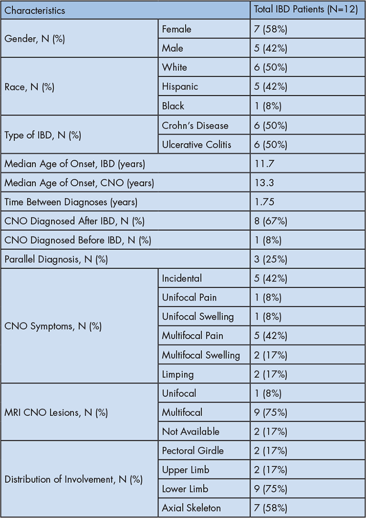
Table 1: Summary of demographic factors, clinical features, and imaging findings in our pediatric inflammatory bowel disease patients diagnosed with chronic nonbacterial osteomyelitisCNO (Chronic Nonbacterial Osteomyelitis), IBD (Inflammatory Bowel Disease)
CNO symptoms varied, with 42% (5 patients) having incidental findings, meaning their CNO was detected without presenting symptoms. Multifocal pain was the most common symptom, reported in 42% (5 patients), followed by multifocal swelling (17%, 2 patients) and limping (17%, 2 patients). Unifocal symptoms were less frequent, with 8% (1 patient) experiencing unifocal pain and another 8% (1 patient) reporting unifocal swelling.
WB-MRI findings showed multifocal lesions in 75% (9 patients), while unifocal lesions were noted in 8%. The most commonly affected regions were the lower limbs (75%, 9 patients) and axial skeleton (58%, 7 patients), followed by the pectoral girdle (17%, 2 patients) and upper limbs (17%, 2 patients).
Lastly, bone biopsy was performed in 17% of the cohort (2 patients); one showed no significant abnormalities, while the other demonstrated non-specific inflammation, edema, and fibrosis
Discussion
This project summarizes our 13-year experience with chronic nonbacterial osteomyelitis in pediatric patients with IBD in South Florida. The demographic distribution varied in our cohort. While European cohorts have shown female predominance1,3,4,14, Latin American and Indian cohorts show male predominance.14 Similarly, our patient population was primarily female. In terms of race, many of our patients identified as White, the group that is most frequently reported to have CNO in previous studies.1,7
In other literature, patients were more likely to have CNO as the initial diagnosis3,7; in our cohort, most patients were diagnosed with CNO after being initially diagnosed with IBD. This may be due to decreased awareness of CNO or possibly the large population of pediatric patients with IBD seen in our institution. CNO remains a rare disease; thus, it is likely underdiagnosed. Patients with IBD are also known to have other joint symptoms such as arthritis; thus, if imaging is not obtained and suspicion for CNO is low, it could go undetected and may underestimate the coexistence rate of both diseases. Our cohort appears to be comparable to the literature-based cohort described by Costi and coworkers, where 17% of their cohort was diagnosed with IBD and CNO simultaneously.3 In our study, 25% of patients had a concurrent diagnosis of both IBD and CNO. This supports the idea that though parallel diagnoses are less common, the index of suspicion must remain high.
Although the etiology of CNO is uncertain, some suggest a link to inflammation in other sites of the body, such as IBD. Many hypotheses have been proposed about the exact cause of the disease, but it has been suggested to involve an imbalance of pro- and anti-inflammatory cytokines.2 Impaired expression of IL-10 and IL-19 has been observed in monocytes from patients with CNO/CRMO.15 Interestingly, altered expression of IL-10 also plays a role in the pathophysiology of IBD. Reduced IL-10 expression due to promoter region polymorphisms is also a proposed pathophysiological mechanism in IBD5, and IL-10-deficient mice have been shown to develop chronic enterocolitis.16 Also, dysregulation of TNF-α, IL-10, IL-12, and IL-23 has been noted in gut immune responses that may affect the joints.7
Dysregulation of gut microbiota has been postulated to lead not only to inflammation in the intestinal environment, but also to inflammation in other areas of the body, such as the bones in CNO and the integumentary system. In patients with CNO, a compromised intestinal barrier could lead to inflammation and the release of cytokines. These cytokines may play a role in triggering extraintestinal manifestations of inflammatory bowel disease.7
Symptoms of CNO vary between patients and may include, but are not limited to, bone/joint pain, swelling, limping, fatigue, or delayed growth.1 CNO can affect multiple bones simultaneously, a phenomenon known as multifocality. Multifocal pain is the most common documented presenting symptom, with other symptoms such as swelling or limping noted less often.3,7 Similar to the current literature, the most common symptom in our patients was multifocal pain, followed by multifocal swelling and limping.
Bone biopsies are sometimes performed early in the diagnostic process, primarily to exclude alternative underlying causes. To date, bone biopsies have not revealed any findings specific or definitive for CNO, which is likely why they are not required for diagnosis. Two of our patients had bone biopsies, and neither biopsy provided diagnostic value in confirming the patients’ diagnosis of CNO. Since no validated diagnostic criteria for CNO currently exist, imaging is vital to the diagnosis. Because WB-MRI remains the gold standard, we diagnosed all our patients based on MRI findings consistent with CNO. Nearly half of patients were asymptomatic and had imaging with incidental findings that prompted further exploration and led to a diagnosis of CNO. In both groups, most patients had multifocal rather than unifocal lesions, consistent with previous literature. Historically, CNO primarily affects the clavicle, vertebrae, mandible, and long-bone metaphyses.1 In our IBD cohort, the most affected bone areas were the lower limbs and axial skeleton.
This case series was conducted at a single institution, focusing on patients diagnosed with IBD who also exhibited radiographic findings indicative of CNO. As a retrospective study, it is subject to selection bias and may lack comprehensive longitudinal data. Since this condition is often underrecognized and requires coexisting IBD and the rarer CNO diagnosis, our sample size was small. Given the absence of validated diagnostic criteria for CNO, we based our assessment on imaging findings, irrespective of whether the patients were symptomatic. Multiple groups have proposed diagnostic criteria/algorithms, but none have been validated to date. The proposed criteria share similarities and encompass a range of major and minor components. These include, but are not limited to, typical clinical and radiographic findings; the coexistence of other autoimmune diseases; the presence or absence of elevated CRP or ESR alongside otherwise normal laboratory results; and a generally good state of health.12-13,17-19 Biopsy findings consistent with inflammatory changes are also considered. We encourage future studies to refine diagnostic criteria, with the goal of developing a validated diagnostic tool for definitive diagnosis.
Lastly, these patients often had poor documentation and follow-up. Steinborn et al. proposed that the first follow-up MRI should be 6 weeks after the start of treatment. In the further course, intervals of 3-6 months would be sufficient. Radiological follow-up is also important after discontinuation of medication to detect early recurrence.20 Due to poor follow-up, we were unable to collect strong data regarding improvement with treatment; thus, treatment response was excluded from our data set. Future studies would benefit from examining the treatments utilized and their corresponding responses
Conclusion
CNO can be considered a rare extraintestinal complication of IBD and should be considered in patients with IBD who experience chronic bone pain or swelling despite unremarkable initial workup. We presented demographic factors, clinical features, and imaging characteristics. Our case series adds to the literature on previously reported cases and further supports the association between CNO and IBD. Raising awareness among pediatricians, hospitalists, rheumatologists, and gastroenterologists about this potential association is crucial to facilitate earlier diagnosis, improve access to treatment, control symptoms, and reduce complications. Importantly, pediatric CNO/CRMO generally has a favorable long-term prognosis. While most patients experience a relapsing-remitting course, the disease typically resolves over time, often without lasting complications. More data are needed to study the clinical and radiographic effectiveness of different treatment modalities.
References
- Zhao DY, McCann L, Hahn G, Hedrich CM. Chronic nonbacterial osteomyelitis (CNO) and chronic recurrent multifocal osteomyelitis (CRMO). J Transl Autoimmun. 2021;4:100095.
- Hofmann SR, Kapplusch F, Girschick HJ, et al. Chronic recurrent multifocal osteomyelitis (CRMO): presentation, pathogenesis, and treatment. Curr Osteoporos Rep. 2017;15(6):542-554.
- Costi S, Germinario S, Pandolfi M, et al. Chronic nonbacterial osteomyelitis and inflammatory bowel disease: a literature review-based cohort. Children (Basel). 2023;10(3):502.
- Reiser C, Klotsche J, Hospach A, et al. First-year follow-up of children with chronic nonbacterial osteomyelitis-an analysis of the German National Pediatric Rheumatologic Database from 2009 to 2018. Arthritis Res Ther. 2021;23(1):281.
- Ramachandran S, Zhao Y, Ferguson PJ. Update on treatment responses and outcome measure development in chronic nonbacterial osteomyelitis. Curr Opin Rheumatol. 2023;35(5):255-264.
- Goldfarb A, Breitling S, Amadasun O, Kiernan B, Gold-von Simson G, Sagalowsky ST. Chronic non-infectious osteomyelitis mimicking scurvy as the presenting sign of Crohn’s disease: case report. Front Pediatr. 2022;10:871993.
- Tzaneti A, Athanasopoulou E, Fessatou S, Fotis L. Chronic nonbacterial osteomyelitis in inflammatory bowel disease. Life (Basel). 2023;13(12):2347.
- Lorenze A, Meadows L, Kehinde T, Menchini CB. A severe presentation of chronic non-infectious osteomyelitis associated with ulcerative colitis: a case report. BMC Pediatr. 2020;20(1):304.
- Nico MAC, Araújo FF, Guimarães JB, et al. Chronic nonbacterial osteomyelitis: the role of whole-body MRI. Insights Imaging. 2022;13(1):149.
- Roberts E, Charras A, Hahn G, Hedrich CM. An improved understanding of pediatric chronic nonbacterial osteomyelitis pathophysiology informs current and future treatment. J Bone Miner Res. 2024;39(11):1523-1538.
- Zhao Y, Dedeoglu F, Ferguson PJ, et al. Physicians’ perspectives on the diagnosis and treatment of chronic nonbacterial osteomyelitis. Int J Rheumatol. 2017;2017:7694942.
- Jansson A, Renner ED, Ramser J, et al. Classification of non-bacterial osteitis: retrospective study of clinical, immunological and genetic aspects in 89 patients. Rheumatology (Oxford). 2007;46(1):154-160.
- Roderick MR, Shah R, Rogers V, Finn A, Ramanan AV. Chronic recurrent multifocal osteomyelitis (CRMO) – advancing the diagnosis. Pediatr Rheumatol. 2016;14:47.
- Koryllou A, Mejbri M, Theodoropoulou K, Hofer M, Carlomagno R. Chronic nonbacterial osteomyelitis in children. Children (Basel). 2021;8(7):551.
- Li MC, He SH. IL-10 and its related cytokines for treatment of inflammatory bowel disease. World J Gastroenterol. 2004;10(5):620-625.
- Marlow GJ, van Gent D, Ferguson LR. Why interleukin-10 supplementation does not work in Crohn’s disease patients. World J Gastroenterol. 2013;19(25):3931-3941.
- Roderick MR, Shah R, Rogers V, Finn A, Ramanan AV. Chronic recurrent multifocal osteomyelitis (CRMO) – advancing the diagnosis. Pediatr Rheumatol Online J. 2016;14(1):47.
- Zhao Y, Wu EY, Oliver MS, et al. Consensus treatment plans for chronic nonbacterial osteomyelitis refractory to nonsteroidal anti-inflammatory drugs and/or with active spinal lesions. Arthritis Care Res (Hoboken). 2018;70(8):1228-1237.
- Nuruzzaman F, Zhao Y, Ferguson PJ. Chronic nonbacterial osteomyelitis: insights into pathogenesis, assessment, and treatment. Rheum Dis Clin North Am. 2021;47(4):691-705.
- Steinborn M, Huf V. Chronic non-bacterial osteomyelitis (CNO) in childhood and adolescence – a disease with many faces. Die Chronische Nicht Bakterielle Osteomyelitis (CNO) im Kindes- und Jugendalter – eine Erkrankung mit vielen Gesichtern. Rofo. 2024;196(3):243-252.